
Bonjour et bienvenue à vous sur Casinogratuits.org qui s’engage à vous dénicher les meilleures informations en matière de casinos avec bonus sans dépôt. Ce site de pari en ligne est dédié à l’activité des casinos en ligne pour tous les francophones. Jour après jour, nous vous fournissons clef en main les informations dont vous avez besoin pour optimiser vos sessions de jeu et même faire de nouvelles découvertes.
Nous savons qu’il s’agit de la promotion la plus recherchée en France. Ainsi, pour vous aider à débusquer la meilleure plateforme de jeu, nous évaluons chaque casino pour francophones jours après jours.
Les 10 meilleurs casinos en ligne bonus sans dépôt
| Classement | Casino | Bonus | NOTE | Visitez |
|---|---|---|---|---|
| 1 |
|
🎁20 Tours Gratuits | VisitezRevue | |
| 2 |
|
🎁20 Tours Gratuits | VisitezRevue | |
| 3 |
|
🎁200% jusqu'à 200€ | VisitezRevue | |
| 4 |
|
🎁100% jusqu'à 500€ | VisitezRevue | |
| 5 |
|
🎁25 Tours Gratuits Book of Dead | VisitezRevue | |
| 6 |
|
🎁20 Tours Gratuits | VisitezRevue | |
| 7 |
|
🎁25 Tours Gratuits | VisitezRevue | |
| 8 |
|
🎁20 Free Spins Offerts Easter Island | VisitezRevue | |
| 9 |
|
🎁20 Free Spins Offerts Easter Island | VisitezRevue | |
| 10 |
|
🎁20 Free Spins Easter Island | VisitezRevue |
Guide rapide des meilleurs jeux de casino gratuits
Le jeu de casino gratuit est une incroyable façon de s’offrir un peu de répit et de prendre du plaisir! Jackpot progressif, bonus sans dépôt, video poker, tout est gratuit est la plupart des jeux sont sans inscription et sans téléchargement. Jouer gratuitement est une merveilleuse façon de se détendre et de peaufiner ses techniques de jeu. Voici les informations essentielles à connaître pour trouver les meilleurs casinos et les meilleurs jeux gratuits.
Nouveaux casinos en ligne : des services plus innovants
Ces 3 dernières années, le secteur des casinos en ligne s’est étoffé de nouveaux acteurs. Très réactifs aux besoins des joueurs, ils proposent de plus en plus de services. L’objectif est d’offrir la meilleure expérience de jeu possible. Pour ce faire, l’accent est mis sur l’innovation et les technologies.
Désormais, les traits communs aux nouveaux casinos en ligne se redessinent, de nouvelles tendances apparaissent. Quelles sont-elles ?
- Dans tous les nouveaux casinos en ligne, l’offre de jeu devient exponentielle. C’est à celui qui offrira le plus de jeux, le plus de machines à sous.
- Tous les nouveaux casinos en ligne proposent des jeux live en direct avec croupier. La présence d’un casino live devient le minimum vital. Ils commencent également à être plus nombreux à proposer des paris sportifs en plus de l’offre pure casino.
- Les bonus et promotions deviennent de plus en plus généreux. Ici, on perçoit des différences sensibles au niveau des wagers associés au bonus. En effet, les casinos sont plus nombreux à proposer des Bonus sans Wager. Ils communiquent fortement dessus et en font un argument de fidélisation. Les bonus casino sans depot deviennent plus fréquents.
- Les cryptomonnaies deviennent la norme. Tous les nouveaux casinos sont des casinos hybrides. Ils acceptent en effet les cryptomonnaies en plus des devises traditionnelles.
Quels sont les nouveaux casinos en ligne ?
AMON
Né en 2023, ce casino profite déjà d’une bonne expérience. En effet, ses concepteurs ont créé le fameux casino Betzino. Il connaît donc bien les habitudes et préférences des joueurs. Son point fort : un bonus sans depot, ce qui se fait de plus en plus rare.
🗓️ Date de création : 2023
💰 Bonus de bienvenue : 200€ + 100 Free Spins
💳 Méthodes de paiement : Cryptos, Visa, MasterCard, Neosurf, CASHlib, Flexepin, eZeeWallet…
💸Dépôt minimum : 20€.
🎰 Jeux : machine à sous, jeux de table, jeux live avec croupier en direct.
📜Fiabilité : licence Curaçao – Cryptage des données
📞Support client : Chat – Email . Support francophone.
GRANDZ CASINO
GrandZ Casino connait depuis peu un grand succès auprès des joueurs. Il est vrai que ce site au design dynamique offre aux joueurs une expérience de jeu exceptionnelle.
🗓️ Date de création : 2023
💰 Bonus de bienvenue : 3000 € + 1000 Free Spins (Pack bienvenue)
💳 Méthodes de paiement : Casino Hybride. Cartes bancaires, Cryptomonnaies… Dépôt minimum : 25€.
🎰 Jeux : machine à sous classique, vidéo, à jackpot, jeux de table, jeux live avec croupier en direct.
📜Fiabilité : licence Curaçao – Cryptage des données
📞Support client : Chat – Email . Support francophone.
RA CASINO
Ra casino est un nouveau casino en ligne qui alligne les performances. Il séduit de nombreux joueurs. Il fait appel à de nombreux fournisseurs de jeux réputés comme Betsoft, ISoftBet, Red Tiger Gaming, Pragmatic Play…
🗓️ Date de création : 2024
💰 Bonus de bienvenue : 100% jusqu’à 500€
📜Fiabilité : licence Curaçao Cryptage des données – Casino sécurisé
💳 Méthodes de paiement : Casino Hybride. Cartes bancaires Visa & Mastercard, ecoPayz, MyFinity, Cryptomonnaies (bitcoin). Dépôt minimum : 50€.
🎰 Jeux : machines à sous classiques, vidéo, à jackpot, jeux de table, jeux live avec croupier en direct/Casino Live.
📞Support client : Chat 24/7 – Email . Support francophone.
JACKPOT BOB
Jackpot Bob accueille les joueurs dans un cadre fiable et sécurisé. Il propose des machines à sous performantes, un large choix de jeux de table comme le poker, la roulette ou le blackjack. Ses nombreux bonus font de ce casino en ligne un lieu attractif fait pour le plaisir du jeu. Il offre en outre un bonus de bienvenue 200% jusqu’à 200€ sans wager.
🗓️ Date de création : 2023
💰 Bonus de bienvenue : 200% jusqu’à 200€ sans wager
📜Fiabilité : licence Curaçao Cryptage des données – Casino sécurisé
💳 Méthodes de paiement : Casino Hybride. Cartes bancaires Visa & Mastercard, ecoPayz, MyFinity, Cryptomonnaies (bitcoin). Dépôt minimum : 20€.
🎰 Jeux : machines à sous classiques, vidéo, à jackpot, jeux de table, jeux live avec croupier en direct/Casino Live.
📞Support client : Chat 24/7 – Email . Support francophone.
Jeux casino gratuit : topo sur les variétés disponibles !
Les jeux de casino gratuits représentent actuellement la quasi-totalité des catégories des jeux en ligne. D’emblée, ils sont regroupés dans le menu principal afin que vous puissiez effectuer votre choix plus facilement. Voici donc l’essentiel de ces jeux :
Les machines à sous
Également connues sous le nom de bandits manchots, il s’agit sans doute des jeux de casino en ligne les plus populaires au monde. Jouer gratuitement sur les machines à sous s’avère intéressant grâce à ses nombreuses lignes de paiement. Vos possibilités de gains seront donc multipliées. Avec le jeu gratuit, vos gains sont bien entendus fictifs, en revanche, les gains vous permettent de jouer plus longtemps.
Jeux de table:
Blackjack : Quel habitué des casinos en ligne ignore l’existence de ce jeu de cartes ? Cependant, gagner une partie de blackjack requiert une certaine maîtrise. En recourant fréquemment aux jeux casino gratuit, vous vous améliorerez à grande vitesse. Le tout est de savoir s’arrêter au bon moment pour surpasser la banque. Jouer aux jeux gratuits de Blackjack permet d’affiner votre instinct de joueur.
| Tableau des jeux de blackjack en ligne les plus populaires | |||
|---|---|---|---|
| Black Russian Blackjack (Ezugi) | Blackjack Platinum VIP (Evolution Gaming) | Classic Speed Blackjack 35 (Evolution Gaming) | Marina Casino Roulette (Ezugi) |
| Blackjack (VivoGaming) | Blackjack Salon Privé (Ezugi) | EMA Black Jack (AsiaGaming) | Power Blackjack (Evolution Gaming) |
| Blackjack (XProGaming) | Blackjack Silver A (Evolution Gaming) | Fiesta Unlimited Blackjack (Ezugi) | Rumba Blackjack (Ezugi) |
| Blackjack Bucuresti (Ezugi) | Blackjack VIP Alpha (Evolution Gaming) | Free Bet Blackjack (Evolution Gaming) | Speed Blackjack M (Evolution Gaming) |
| Blackjack Diamond VIP (Evolution Gaming) | Blackjack VIP Beta (Evolution Gaming) | Infinite Blackjack (Evolution Gaming) | Speed VIP Blackjack A (Evolution Gaming) |
| Blackjack Fortune VIP (Evolution Gaming) | Blackjack VIP Gamma (Evolution Gaming) | Italian Blackjack (Ezugi) | Turkish Blackjack (Ezugi) |
| Blackjack Gold 6 (Ezugi) | Blackjack VIP Z (Evolution Gaming) | J1 Blackjack (HoGaming) | Unlimited Blackjack (Ezugi) |
| Blackjack Grand VIP (Evolution Gaming) | Burgas Blackjack (VivoGaming) | J2 Blackjack (HoGaming) | VIP Blackjack (Ezugi) |
| Blackjack Party (Evolution Gaming) | CA1 Blackjack (HoGaming) | Lightning Blackjack (Evolution Gaming) | VIP Blackjack with Surrender (Ezugi) |
| Blackjack Platinum (Ezugi) | Classic Speed Blackjack 12 (Evolution Gaming) | Mambo Unlimited Blackjack (Ezugi) | White Russian Blackjack (Ezugi) |
Baccarat:Joueurs avides de sensations fortes, le Baccarat est fait pour vous. Les jeux free de casino vous permettent d’apprécier un jeu similaire à la roulette, avec un rythme plus rapide.
Roulette: Vedette des jeux de table, la roulette combine simplicité et habileté. Toutefois, ce jeu d’argent basé sur le hasard peut être maîtrisé à travers certaines stratégies bien posées. Tout type de joueur aura la chance de rafler la mise s’il s’entraîne régulièrement sur le mode démo.
| Tableau des jeux de roulette en ligne les plus populaires | |||
|---|---|---|---|
| American Roulette (Evolution Gaming) | French Roulette Gold (Evolution Gaming) | Onyx Roulette (EGT interactive) | Roulette VIP (LiveGames) |
| Automatic Roulette (Ezugi) | Grand Casino Roulette (Evolution Gaming) | Oracle 360 Roulette (Ezugi) | Ruleta del Sol (Ezugi) |
| Auto-Roulette La Partage (Evolution Gaming) | Hippodrome Grand Casino (Evolution Gaming) | Portomaso Roulette (Ezugi) | Ruletka Russia (Ezugi) |
| Auto-Roulette VIP (Evolution Gaming) | Immersive Roulette (Evolution Gaming) | Roulette (VivoGaming) | Speed Auto Roulette (Evolution Gaming) |
| Casino Malta Roulette (Evolution Gaming) | Instant Roulette (Evolution Gaming) | Roulette (XProGaming) | Speed Auto Roulette (Ezugi) |
| Cumbia Ruleta (Ezugi) | Italian Roulette (Ezugi) | Roulette Live (HoGaming) | Speed Roulette (Evolution Gaming) |
| Diamong Roulette (Ezugi) | Lightning Roulette (Evolution Gaming) | Roulette Lobby (Ezugi) | Speed Roulette (Ezugi) |
| Double Ball Roulette (Evolution Gaming) | Live European Roulette (EGT interactive) | Roulette of AGIN (AsiaGaming) | Türkçe Rulet (Ezugi) |
| Dynamic Roulette 120x (EGT interactive) | Live Speed Roulette (EGT interactive) | Roulette of AGQ (AsiaGaming) | VIP Roulette (Evolution Gaming) |
| Fiesta Roulette (Ezugi) | Marina Casino Roulette (Ezugi) | Roulette Speed (LiveGames) | Vivo Gaming Lobby (VivoGaming) |
Video Poker: Le mariage parfait entre la machine à sous et le Poker. Il faut être un bon stratège pour sortir victorieux d’une partie de vidéo poker. Néanmoins, il importe d’aiguiser ces compétences pour assurer autant que possible un gain répétitif.
Keno: la version en ligne des jeux de hasard avec tirage de numéros. Il vous suffit de trouver la combinaison gagnante, ce qui ne vous empêche pas de vous entraîner au préalable. De nombreux casinos en ligne proposent la version démo du Keno.
Poker: Les jeux de poker en ligne offrent des alternatives intéressantes et permettent de vous amuser sans vous ruiner. En guise de proposition, jouez gratuitement sur Texas Hold’em Poker.
Jeux de grattage :
La procédure se limite à gratter les symboles à l’aide de votre souris. Cependant, ne vous précipitez pas à y miser votre argent. Il est plus prudent de tenter votre chance avec les jeux gratuits de casino. Vous pourrez ainsi jouer sans dépenser.
Craps: Les casinos en ligne reprennent également ce jeu de dés pour vous divertir davantage. Avec la version démo, vous saurez au préalable ce que la chance vous réserve.
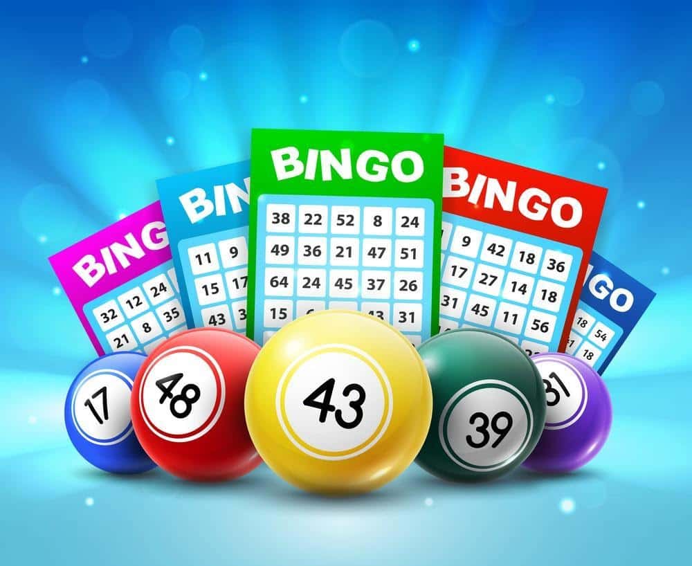
Bingo: Les jeux de casino donnent un coup de jeune au célèbre bingo et vous invitent à le découvrir pour sa version en ligne. Et mieux encore, divertissez-vous avec le mode démo.
Sic Bo: Ce jeu originaire de Chine vous donne la chance de parier avec le tirage de 3 dés. Pour maximiser vos gains ou profiter tout simplement des moments de détente, optez pour la version démo du Sic Bo.
Comment utiliser le guide des meilleurs jeux de casino?
Ce guide rapide des meilleurs jeux de casino a pour vocation d’améliorer l’expérience de jeu sur les casinos en ligne bonus sans dépôt. C’est pourquoi, nous réalisons des examens réguliers des jeux de casino et des établissements qui en proposent. Nous vous proposons un tas d’astuces pour tester les jeux.
Parmi elles, il existe le mode démo, disponible sur un bon nombre de jeux de casino. Les joueurs peuvent, grâce à ce mode, jouer juste pour le fun aussi longtemps qu’ils le souhaitent.
Les bonus sans dépôt sont aussi très intéressants pour tester de véritables jeux avec de l’argent casino offert sans obligation de dépôt. Pour en profiter il vous suffit de créer un compte sur un casino en ligne bonus sans dépôt et de réclamer votre bonus!
Vous trouverez une grande quantité de bons bonus sans dépôt dans notre classement des meilleurs casinos en ligne. N’hésitez pas à consulter nos revues, elles comportent souvent des codes exclusifs uniquement dédiés à nos lecteurs!

Quels sont les avantages des jeux de casino gratuits?
Cette question vous a sûrement traversé l’esprit à un moment donné. Les bénéfices sont nombreux : par exemple, les jeux gratuits de casino ne requièrent aucun engagement préalable. De plus, ils vous permettent de vous familiariser avec votre jeu favori avant de vous lancer dans les mises en argent réel. Vous pouvez alors vous divertir tout en restant informé des dernières tendances. Une fois que vous jouez en mode réel, vous y gagnerez peut-être une coquette somme. Cela avec l’aide du talent, de la providence et de l’expérience sur les jeux de casino gratuit bien sûr.
Comment gagner dans un casino en ligne bonus sans dépôt ?
Tout le monde joue dans l’espoir de gagner, tout en profitant de chaque moment de la partie. Ces conseils vous seront d’une aide précieuse dans votre quête de victoire. En les appliquant dans les jeux gratuits, vos chances de gains augmenteront certainement. En plus, dans un casino en ligne bonus sans dépôt, il est possible de gagner et de retirer les gains réalisés grâce eux. D’où l’intérêt des bonus sans depot !

Premièrement, il vous faut maîtriser parfaitement les règles du jeu. Au lieu de perdre de l’argent en vous reposant sur la chance, jouez de manière intelligente. Familiarisez-vous avec les jeux gratuits de casino avant de jouer de l’argent réel.
Deuxièmement, engrangez de l’expérience. Afin de retenir l’attention des joueurs, les développeurs renouvellent constamment leurs titres. Par conséquent, de nombreuses heures d’entraînement vous seront sans doute nécessaires avant d’acquérir toutes les ficelles du jeu.
Troisièmement, pensez à établir une stratégie mathématique. Les jeux de hasard sont soumis aux principes de la probabilité. Aussi, le fait de pouvoir identifier les tendances naturelles et à miser en conséquence permet d’augmenter les gains. Enfin, faites preuve de discipline et surtout amusez-vous!
Les casinos avec bonus sans dépôt made in France

Quoi de plus simple que de trouver des plateformes de casino sur internet ? Mais avez-vous la certitude que cette plateforme est fiable ? L’important c’est de savoir sur quelle plateforme il est bon d’éviter de mettre les pieds ! De plus l’ANJ (Autorité Nationale des Jeux) a mis en place en 2010, une loi qui complique l’existence et le bon fonctionnement des casinos en France. Depuis, beaucoup d’entre eux ont dû revoir leurs ambitions d’offres à la baisse voire même, mettre la clef sous la porte. Certes, cela a permis de se débarrasser des sites frauduleux. Mais cela a aussi empêché les Français de profiter de casino en ligne bonus sans dépôt de bonne réputation. Ces derniers ont préféré renoncer au marché français à la législation très contraignante.
Alors trouver une plateforme française qui correspond à vos besoins se révèle parfois être une tâche ardue. C’est pourtant essentiel de ne pas se tromper. Casinogratuits.org vous indique quelles plateformes sont les plus fiables avec un support francophone et avec les meilleures offres.
Des informations sur les concepteurs
Outre les casinos avec bonus sans dépôt, nous vous fournissons de nombreuses informations sur les concepteurs de jeux de machines à sous vidéo. Leur réputation est la garantie d’une session de jeu de qualité. Des graphismes de nouvelle génération et des effets 3D bluffants, qui s’apparentent presque aux jeux vidéo. Découvrez à travers nos revues, quelles machines sont susceptibles de vous apporter le plus de satisfaction. Betsoft, NetEnt, Isoftbet, RTG, certains d’entre eux fournissent près de 500 casinos dans le monde comme Betsoft. Certains autres vont d’avantage mettre en avant leurs points forts. Mettre en vedette plus de 150 machines à sous et les plus gros jackpots progressifs comme c’est le cas pour RTG. En outre, la rédaction fournit son expertise sur ces développeurs et vous renseigne sur leur qualité. Voici comment tout savoir sur les casinos avec bonus sans dépôt.
Voici une liste des concepteurs de jeux fiables par ordre alphabétique ayant la plus grande notoriété sur le marché des casinos en ligne :
| Tableau des concepteurs de jeux en ligne fiables | |||
|---|---|---|---|
| AvatarUX Studios | Endorphina | NetEnt | Red Rake Gaming |
| Betconstruct | EvoPlay | Nolimit City | Red Tiger Gaming |
| Betgames | FIAT | OneTouch | Rival Gaming |
| Betsoft | Fugaso | Pari Play | Spinomeal |
| Betgames | Gameart | Platipus Gaming | Thunderkick |
| bGaming | Ganapati | Play’n GO | Vivo Gaming |
| Big time gaming | High 5 Games | Playson | Xplosive |
| Booming Games | Igt | Playtech | Yggdrasil |
| Boongo | Iron Dog Studio | Pragmatic Play | 1X2 Gaming |
| DreamTime Gaming | Kalamba Games | Quickspin | 2by2 Gaming |
| ELK | Microgaming | Rabact | 4theplayer.com |
Casino en ligne gratuit

Être membre de nos lecteurs assidu c’est aussi profiter de nos codes promo exclusifs qui vous donnent accès aux casinos avec bonus sans dépôt. En tant que joueur, il est parfois important de dénicher ces casinos avec bonus sans dépôt pour pouvoir s’acclimater aux plateformes de jeux et établir vos stratégies de jeu.
Casino en ligne bonus sans dépôt : Le jeu gratuit
Certains se demandent souvent pourquoi les joueurs de casino en ligne sont à la recherche de plateformes gratuites ? La raison est simple. Premièrement, il est plus aisé de s’entrainer sur ce type de casinos avant d’y engager son propre argent si durement acquis. De plus, sur un casino en ligne bonus sans dépôt, l’offre de jeu gratuite n’est limitée ni par le temps ou ni par le nombre de tours. Aux yeux d’un joueur en cours de perfectionnement, les casinos en ligne gratuits lui confèrent un élément essentiel pour devenir un « tueur » dans l’univers du casino en ligne, le temps.
De plus ce type de plateforme propose des avantages intéressants bien différents de ceux que proposent le casino en ligne sans dépôt. Le service offert par ce type de casino promet également une approche plus didactique et ludique que le casino avec bonus sans dépôt, s’il s’agit seulement de faire passer le temps.
Casino avec bonus sans dépôt : comment débuter ?
S’inscrire sur un casino avec bonus sans dépôt, c’est être prêt à plonger dans le grand bain. En effet, le casino avec bonus sans dépôt est à la base une plateforme qui requière un investissement en argent réel de la part du joueur. Dans le but de faciliter votre transition vers le jeu en argent réel, le casino avec bonus sans dépôt peut mettre à votre disposition ce type d’offre.
Casinos avec bonus sans dépôt : En quoi ça consiste?

Un bonus sans dépôt est une offre promotionnelle dédiée généralement aux nouveaux joueurs. Cette promotion a pour vocation de souhaiter la bienvenue à ses nouveaux membres. Les casinos avec bonus sans dépôt peuvent offrir aux joueurs de l’argent, des tours gratuits (free spins), du cashback. Notre site vous aidera à identifier quel bonus est le plus adapté pour votre jeu et quel est le meilleur bonus casino sans depot.
Si vous êtes un fan de bandit manchot, ca tombe bien ! La majorité des bonus sont spécialement dédiés aux machines à sous. Le bonus vous aidera à en tester un plus grand nombre. Cependant, les autres jeux ne sont pas obligatoirement exclus.
Si vous êtes plutôt blackjack, roulette ou encore poker, pensez à vous mettre en quête du bonus utilisable aussi sur ces jeux. Certains bonus ont des restrictions d’utilisation très strictes. Certains casino en ligne bonus sans dépôt ont des politiques très strictes. Ils suppriment votre bonus destiné initialement aux machines à sous si vous en faites usage sur une table de jeu. Pensez à consulter minutieusement les conditions d’utilisation !
Notre objectif est de vous guider de la façon la plus honnête possible pour vous permettre de jouer sereinement et en pleine connaissance de cause. Nous mettons à votre disposition des revues analysant les plateformes sur lesquelles vous souhaitez potentiellement parier ; mais aussi des articles vous informant des dernières nouveautés sur le jeu en ligne. Quels sont les pièges à éviter ? servez-vous de notre expérience pour optimiser votre expérience de jeu.
Les 10 meilleurs casinos bonus sans depot 2024
Un casino bonus sans depot offre le petit plus dont tous les jours raffolent : la possibilité de jouer gratuitement! Quoi de mieux pour tester les offres et performances d’un casino en ligne qu’un bonus sans depot ? Rien, justement. Malheureusement, ces promotions se font de plus en plus rares sur internet. Mais il est encore possible d’en trouver en cherchant bien. Nous sommes parvenus à établir une liste des 10 meilleurs casinos 2024 proposant les meilleurs bonus casino sans depot.
| Bonus Sans Depot | Bonus | Bonus Max | Free Spins | Wager | Retrait Max. Hebdo. | Retrait Max. Mensuel | |
|---|---|---|---|---|---|---|---|
| Cresus | 20 FS | 150% | 300€ | 600 | - | 2 500€ | 10 000€ |
| Lucky 8 | 20 FS | 100% | 200€ | 500 | x40 | 2 500€ | 10 000€ |
| Ra Casino | 20 FS | 100% | 500€ | - | x40 | 1 500€ | 6 000€ |
| Amon Casino | 25 FS | 150% | 200€ | - | x40 | 1 500€ | 6 000€ |
| Lucky31 | 20 FS | 100% | 150€ | - | x30 | 5 000€ | 20 000€ |
| Betzino | 25 FS | 100% | 200€ | 100 | x30 | 5 000€ | 20 000€ |
| Extra Casino | 20 FS | 100% | 50€ | 100 | x30 | 5000€ | 20 000€ |
| DublinBet | 20 FS | 100% | 100€ | - | x30 | 2 500€ | 5 000€ |
| WinOui | - | 100% | 1 000€ | 300 | x30 | 2 500€ | 10 000€ |
| Tortuga | - | 120% | 1 200€ | - | x40 | 2 500€ | 10 000€ |
● Cresus Casino

Commençons notre top 10 des meilleurs casinos 2024 en beauté avec Cresus. Véritable référence sur la scène francophone, ce casino en ligne vous promet des performances de haut vol. Il vous donne accès aux plus grands succès des fournisseurs de jeux les plus réputés. Mais ce n’est pas tout ! Ses prestations sortent également du lot. Vous le comprendrez au vu des offres exclusives et des promotions tentantes. Et pour couronner le tout, un service client efficace est à votre entière disposition. Il est à votre écoute et répond à toutes les questions. Comment annuler un bonus actif Cresus Casino ? Comment fonctionne un bonus au Casino ? Ils savent vous apporter toutes les informations nécessaires. N’hésitez plus et inscrivez-vous ! Vous aurez l’opportunité de découvrir gratuitement les offres grâce au bonus sans depot. Celui-ci vous donne droit à 20 Free spins sur Multifly d’Yggdrasil.
● Lucky 8

Continuons notre classement des 10 meilleurs casinos 2024 offrant des bonus sans depot avec Lucky8. Ce casino en ligne se démarque clairement du lot avec un site très bien fait. Le design est soigné et facilite la navigation. Par ailleurs, les prestations et performances se montrent à la hauteur de ce que l’on attend d’un bon casino. Pour en savoir un peu plus sur ses offres promotionnelles et ses bonus exclusifs, pourquoi ne pas essayer le bonus sans depot ? Actuellement, Lucky8 offre un bonus sans depot de 20 tours gratuits. Ils vous permettront de tester le site et surtout de bien en profiter.
● Amon Casino

Pour une expérience de jeu inégalée, vous pourrez toujours compter sur Unique Casino. Ce site mise sur des services et prestations hors du commun pour vous satisfaire. Sa large gamme de jeux comblera les joueurs et les parieurs chevronnés. Ses promotions et bonus constituent également des occasions rentables indéniables. Ses modes de paiement divers et variés se veulent pratiques pour le retrait des gains. Ce n’est donc pas étonnant si l’établissement intègre notre top 10 meilleurs casino bonus sans depot 2024. D’ailleurs, son bonus sans depot de 25 free spins prouve sa détermination de vous séduire et de vous surprendre.
● Casino Extra

Que serait un classement des 10 meilleurs casinos 2024 sans Casino Extra mérite sans conteste sa place dans ce classement des 10 meilleurs casino bonus sans depot ? Ce casino en ligne jouit d’une réputation sans faille sur la scène française pour plusieurs raisons. Une ribambelle de promotions alléchantes sont conçues pour pimenter vos sessions de jeu. Bonus de bienvenue, cashback, … rien n’est laissé au hasard. En outre, le bonus sans depot accessible à l’inscription est à tester sur le slot Spina Colada™ d’Yggdrasil.
● Betzino

Poursuivons notre liste des 10 meilleurs casinos 2024 avec Betzino. Les joueurs français apprécient ce casino en ligne qui propose des services impécables. Le catalogue de jeux intégrant les meilleures machines à sous et un excellent casino live est un avantage ! Alimentée par les plus grands fournisseurs de jeux au monde, celle-ci vous garantit de belles sessions de machines à sous, jeux de table, etc. Pour découvrir les offres de Betzino, réclamez votre bonus sans depot. Ainsi, vous profiterez de 25 tours gratuits sur Book of Dead.
● Ra Casino

Ra Casino tient une bonne place dans ce classement des 10 meilleurs casinos 2024. Son catalogue de jeux, équipée des meilleurs logiciels, convainc de nombreux joueurs. Par ailleurs, ses promotions hebdomadaires et ses bonus représentent des occasions rentables à ne pas manquer. Pour tester gratuitement les performances de ce casino en ligne, vous avez la possibilité de créer un compte utilisateur. En réclamant votre bonus sans depot, 20 free spins vous seront offerts.
● Lucky31

Amusez-vous sur un casino en ligne fiable et sécurisé en vous connectant sur Lucky31. Différents modes de paiement, dont les crypto-monnaies, sont prévus pour votre confort d’utilisation. De plus, des centaines de titres développés par les meilleurs studios vous donneront l’opportunité de vous adonner à votre passion du pari. Si vous vous inscrivez sur cet établissement, vous serez en droit de demander un bonus sans depot exclusif. En effet, 20 free spins sur toutes les machines à sous vous permettront de jauger des performances du site.
● Tortuga Casino

Pour vous immerger dans un univers original et attractif, rien de tel qu’une partie de machine à sous sur Tortuga. Grâce à son concept sur le corsaire des mers, le Capitaine Tortuga, ce casino en ligne s’est bâti une solide réputation. Par ailleurs, sa multitude d’offres exclusives vous fera saliver. Bonus hebdomadaires, tournois exclusifs, bonus sans depot ainsi que promotions en tout genre sauront vous surprendre.
● DublinBet

DublinBet met à votre disposition un site sobre dédié aux paris en argent réel. Ce casino en ligne s’illustre par son catalogue de jeux hors normes de machines à sous, casino live et jeux de table. Si vous souhaitez vous enquérir de ses promotions et prestations, pourquoi ne pas créer votre propre compte ? Le bonus sans depot associé vous attribue 20 Free Spins sur Holmes and the Stolen Stones d’Yggdrasil.
● Winoui

Pour terminer notre classement des 10 meilleurs casinos 2024, intéressons-nous à Winoui. Ce casino en ligne au design élégamment sobre séduit par la qualité de sa ludothèque. De plus, des bonus et promotions exclusives vous permettront d’assouvir votre soif de pari dans les meilleures conditions. Winoui propose épisodiquement des bonus sans depot à ses joueurs. Le plus souvent, il s’agit de free spins.
Pour vous aider dans votre quête d’opportunités rentables et de fun, nous vous avons listé les 10 meilleurs casinos bonus sans depot 2024. Outre leurs offres intéressantes, ces casinos se distinguent aussi par leur fiabilité.
Le guide du débutant
Faire son entrée dans un nouvel environnement de jeu nécessite d’en apprendre les règles. Certaines têtes brûlées de joueurs foncent parfois dans le monde des casino en ligne bonus sans depot, tête baissée avant de les apprendre dans la douleur.
C’est donc avec bienveillance, que nous vous livrons les « trucs et astuces » qu’un joueur débutant doit connaitre.
Leçon n°1 : Ne jamais négliger les vertus de l’entraînement sur un casino bonus sans depot

Nous le répétons assez souvent, il n’y a rien de pire que de perdre son argent à un jeu que l’on ne comprend pas. C’est pourquoi, un casino bonus sans depot est votre meilleurs outils pour créer vos tactiques de jeu. Grâce à lui, vous serez paré à quasiment tous les cas de figure. Les joueurs les plus méthodiques établissent même un tableau de stratégie. Cela leur permet de ne pas faire d’erreur lorsqu’ils sont confrontés à un choix de coup compliqué dans un temps de jeu restreint. Les moments d’hésitation conduisent parfois à commettre des erreurs. Ce tableau de stratégie recense toutes les situations auxquelles le joueur a été confronté. En cas d’hésitation, un léger coup d’œil à votre anti-sèche personnalisée ne fera pas de mal à votre style de jeu. Surtout si vous jouez de chez vous sur des casinos avec bonus sans dépôt !
Une fois votre analyse de chaque étape de jeu faite et que vous vous estimez prêt, lancez-vous! Il est vrai que les jeux gratuits, au bout d’un moment, c’est ennuyeux. Ils n’ont pas les mêmes propriétés d’attraction que les jeux d’argent. Jouer avec la sueur de votre front rend l’enjeu plus important. Chaque coup comporte son lot d’adrénaline et c’est bien ce que les joueurs de grosses sommes d’argent recherchent. Mais attention, ce sentiment est addictif.
Leçon n°2 : Se fixer un budget
Avant d’entrer dans l’arène, il faut d’abord savoir se dompter soi-même. Pour cela soyez discipliné, cela vous sauvera la mise à plusieurs reprises. Pour commencer, prévoyez avant tout un budget entièrement dédié au casino en ligne. Vous aurez déjà une idée de la limite financière à ne pas franchir. Pour établir ce budget, rien de plus simple. Calculez d’abord vos dépenses fixes, les charges pour votre habitation, l’argent à dépenser dans les transports, les dépenses vitales et la nourriture pour le chat. Il est important que le budget « jeux » n’empiète pas sur vos dépenses vitales. Puis établissez un budget loisir. Toutefois, il doit contenir, vos vacances, petits week-ends en amoureux et autres activités liées à la détente. Dans ce budget, définissez une partie dédiée aux casinos avec bonus sans dépôt.
Leçon n°3 : Respectez les limites fixées !
Tout d’abord, nous le savons mieux que personne, le gain est parfois tentant. Qui n’à pas eu envie de rejouer pour continuer à gagner ? Mais il faut savoir s’imposer des limites et ne pas se laisser déborder par l’euphorie du jeu ! Par conséquent, il est important de rester fidèle à la limite que vous vous êtes fixé. Sachez vous arrêter au bon moment.
Leçon n°4 : Le choix du casino bonus sans depot

Puisque jouer en toute confiance, est essentiel. Il faut donc bien choisir son casino. Il s’agit de loin du choix le plus crucial à faire pour un nouveau joueur. Pour ce faire, une petite étude préalable est de mise. Basez-vous premièrement sur la licence que possède votre plateforme de jeu. En l’absence de licence, abstenez-vous. La licence est pour un casino en ligne un gage de sécurité et de fiabilité. A l’image d’un contrat de travail pour l’employeur, le casino en ligne s’engage à respecter les règles en échange de cette licence. Ce document vous garantie que la plateforme est règlementée et respecte les règles.
Evidemment, certaines licences sont plus faciles à obtenir que d’autres. Les licences les plus cotées proviennent d’Europe : de Malte, Gibraltar, de Chypre et du Royaume-Unis. Cependant, les nouvelles plateformes optent généralement pour une licence délivrée par l’Etat de Curaçao situé dans les Caraïbes ou du Costa Rica. Bien que plus faciles à obtenir que les licences européennes, ces licences sont aussi respectables. Elle sont une preuve de légalité de votre plateforme de jeu.
Les réglementations
Les plateformes européennes sont contrôlées par l’ANJ (anciennement l’ARJEL). C’est pourquoi en tant que joueur français, elles sont à privilégier. En cas de litige, vous pourrez toujours faire jouer la régulation Européenne.
Ces casinos en ligne sont soumis à des règles plus strictes. Cependant, les licences délivrées par d’autres pays le sont aussi.
Afin d’avoir une vision plus claire des plateformes sûres, nous mettons à votre disposition un classement des meilleurs casinos en ligne. Etudiés au peigne fin, vous pourrez bénéficier de toutes les informations relatives à la plateforme de jeu selon celle qui suscite votre attention. Et ainsi en connaître les avantages et les inconvénients.
Vous trouverez ci-dessous les licences de jeu les plus connues et les plus sûres que vous trouverez sur les casino bonus sans depot. Vous pouvez vous y fier.
| Licences des casinos en ligne | |
|---|---|
 | Autorité des Jeux en ligne de Curaçao |
 | Autorité des Jeux de Malte |
 | Commission des Jeux de Kahnawake |
 | Autorité de Régulation de Gibraltar |
 | Autorité National des Jeux |
 | Commission des Jeux de Hasard de Belgique |
 | Commission Fédérale des Maisons de Jeu |
Leçon n°5 : Le service assistance client dans un casino bonus sans dépôt

Puis, il est important qu’au cours de vos sessions de jeu, vous puissiez obtenir des informations ou de l’aide dans les plus courts délais. C’est pourquoi la qualité du service assistance client est un facteur déterminant. Premièrement, il est toujours plus rassurant d’avoir affaire à des opérateurs qui s’expriment dans une langue que vous comprenez. Ensuite, veilliez à ce qu’ils puissent être disponibles par plusieurs canaux de communication. De plus, de nombreux nouveaux casinos en ligne proposent un chat live. En effet, les joueurs apprécient particulièrement cet espace de discussion ou les opérateurs répondent en temps réel.
De plus, les plateformes de jeu rédigent souvent une page FAQ qui pourra grandement vous renseigner sur de nombreux sujet. Comment fonctionne un bonus sans depot ? Comment annuler bonus actif ? Le service client a pour vocation de vous rassurer et de vous informer.
Les 4 options pour l’assistance des casinos en ligne :
- Chat en direct (recommandé) 💬
- Appel téléphonique ☎
- Email 📧
- FAQ ❔
Petit plus : il semblerait qu’une bonne relation avec le service client peut vous permettre de profiter de petites cadeaux comme des bonus sans depot, du cashback ou des free spins.
Leçon n°6 : Les casinos avec bonus sans dépôt
Ils sont de plus en plus nombreux à multiplier leurs offres de bonus sans dépôt. Il est vrai qu’un bonus sans depot est un argument de poids pour attirer les joueurs. Et pour vous arracher à la concurrence, le marché du casino en ligne sait être généreux. Ainsi, il vous attire à coup de bonus aguicheur, de tours gratuits, de free spins. Un casino bonus sans depot peut aussi offrir du temps de jeux gratuit, des jetons cadeau ou bonus de recharge.
La concurrence, c’est une aubaine pour les joueurs qui, par conséquent, peuvent bénéficier d’une multitude d’offres promotionnelles parfois très avantageuses. Le joueur fait sont marché et choisi les offres les plus généreuses. Il change de casino en fonction des bonus offerts. S’inscrit sur un casino bonus sans depot pour profiter d’une nouvelle promotion. Mais attention les casinos avec bonus sans dépôt ont aussi des règles strictes qu’il faut connaître :
- Les bonus sont soumis à des wagers, des conditions de mises strices.
- L’argent offert ne peut pas toujours être retiré facilement en argent réel
- Certains bonus ne sont pas utilisables sur tous les jeux.
- Le bonus de bienvenue n’est utilisable qu’une seule fois par le joueur seulement après inscription.
Les bonus de bienvenue ont pour but de vous laisser tester les jeux de la plateforme. De vous faire une idée de quelles machines vous plaisent le plus et vous inciteront à parier.
Ces bonus ne sont pas les plus avantageux, mais pour un joueur débutant, cela peut se révéler un excellent moyen de jauger une plateforme.
Mais ne soyons pas dupes. Un casino bonus sans depot n’est pas une entreprise philanthropique. Le but est de vous amener à parier avec votre propre argent. C’est aussi un business.
Leçon n°7 : Le bonus avec dépôt
Les bonus avec dépôt sont en général les plus intéressants. Habituellement présentés sous forme de pourcentage, ces bonus peuvent varier entre 100% et 400% sur un premier dépôt. Et ces offres sont parfois limitées à un dépôt d’une somme maximale. Par exemple, si vous recevez un bonus de 100% dans la limite des 150 euros de dépôt. Cela veut dire que lorsque vous effectuez un dépôt de 150 euros sur votre compte joueur, puis vous recevez 150 euros de bonus offert par le casino. Les mises varient ensuite en fonction de votre expérience et de votre fréquence de jeu.
Leçon n°8 : Les moyens de paiement
Enfin, plusieurs moyens de paiement sont proposés aux joueurs pour assurer la protection de leurs données bancaires. De plus, certains moyens de paiement alternatifs comme les portefeuilles électroniques. Avec l’apparition des casinos bitcoin, l’utilisation des cryptomonnaies s’est généralisée en tant que mode de paiement. Désormais, en plus des bitcoin casino, coexiste des casinos hybrides acceptant les devises classiques et les cryptomonnaies.
Aujourd’hui, dans les nouveaux casinos en ligne ou bonus casino sans depot, les mode de versements sont multiples. Les dépôts peuvent être notamment effectués par carte bancaire, par chèque, carte prépayée, et portefeuilles électroniques de type : Neteller, Moneybookers, Ecocard, cryptos etc…
Cependant, n’hésitez pas à vous renseigner auprès du service assistance client de la plateforme de votre choix pour obtenir des informations complémentaires.
Comment déposer sur un casino en ligne ?
| Méthodes pour déposer sur un casino en ligne | ||
|---|---|---|
| Carte bancaire | Visa, Visa Pro, Mastercard, Maestro | Instantané |
| Cryptomonnaies | Bitcoin, Bitcoin Cash, Dogecoin, Ethereum, Litecoin, Tron, XRP... | Instantané |
| E-wallets | Cashlib, Muchbetter, Neosurf, Neteller, Paysafecard, Skrill... | Instantané |
| Virement bancaire | Banque | 2 – 3 jours |
En bref, en ce qui concerne les casinos avec bonus sans dépôt, vous connaissez maintenant le B-A BA du casino en ligne pour débutant. En conclusion, mesdames et messieurs, faites vos jeux !
✨Qu'est-ce qu'un bonus sans depot?
Un bonus sans depot est un cadeau du casino permettant au joueur de jouer gratuitement, sans déposer d'argent. Cela lui permet de tester le casino en ligne bonus sans depot.
✨Comment annuler bonus actif Cresus Casino
Il est possible d'annuler un bonus actif Cresus Casino en le demandant au service client de Cresus Casino.
✨Quel casino offre bonus sans depot ?
De nombreux casinos offrent des bonus sans depot. On les désigne désormais sous l'appellation casino bonus sans depot ou encore casino en ligne bonus sans dépôt. Vous trouverez sur notre site les meilleurs casino bonus sans depot et leurs offres.
✨Comment contacter un service client ?
Un bon casino en ligne bonus sans dépôt propose à ses joueurs des support clients efficaces qu'ils peuvent contacter de plusieurs façons.
1- Live Chat
2- Mail
3- Téléphone (pour certains)
✨Qu'est-ce qu'un casino Hybride ?
Un casino hybride est un établissement acceptant à la fois les devises traditionnelles, mais aussi les cryptomonnaies.
✨Comment fonctionne un bonus au casino ?
Lorsqu'on vous accorde un bonus, vous avez aussitôt droit de l'utiliser. Il peut s'agir de tours gratuits (Free Spins), d'argent, de cashback, de temps de jeu gratuit.... En fonction des casinos en ligne bonus sans depot, il faut soit les activer directement sur le site, soit passer par le service client. Dès qu'ils apparaissent sur votre compte, vous pouvez commencer à jouer.
✨Quels sont les avantages des cryptomonnaies dans des casinos en ligne ?
Jouer dans un casino bitcoin ou un casino hybride comporte de nombreux avantages.
1- Des bonus plus généreux.
2- Une plus grande confidentialité des transactions.
3- Des délais de retraits instantanés.